As published in Fertility and Sterility Vol. 65, No. 6, pp. 1119-1124
Elizabeth A. Stewart, M.D.,
Douglas J. Austin, M.D., Prachee Jain, B.A., Martha D. Penglase, A.B., Romana A.
Nowak, Ph.D.
From the Department of Obstetrics,
Gynecology and Reproductive Biology, Harvard Medical School, Brigham and Women's
Hospital, Boston, Massachusetts
Objective: To assess the action of RU486 (mifepristone),
in the presence and absence of P, on PRL production by explant cultures of
leiomyoma and myometrium.
Design: Explant cultures using tissue from nine premenopausal women
undergoing hysterectomy in the proliferative phase of the menstrual cycle;
immunohistochemical staining of tissue sections from five patients for P
receptor (PR) subtype.
Main Outcome Measures: Prolactin secretion (measured by RIA), lactate
dehydrogenase secretion (measured by quantitative colorimetric assay), and
immunohistochemistry for PR subtype.
Results: Prolactin secretion was decreased in leiomyomas by RU486 at
concentrations of 10-7 M and 10-5 M when normal
serum-containing medium was used. In experiments with all detectable P removed
from serum, PRL secretion was suppressed in both leiomyomas and myometrium at an
RU486 concentration of 10-7 M. Immunohistochemistry results suggest
that the A form of the PR is the dominant form in both leiomyomas and myometrium.
Conclusions: Prolactin production is suppressed in both leiomyomas and
myometrium after treatment with RU486 in vitro, and this suppression may serve
as a marker for the clinical effectiveness of agents used in the treatment of
leiomyomas.
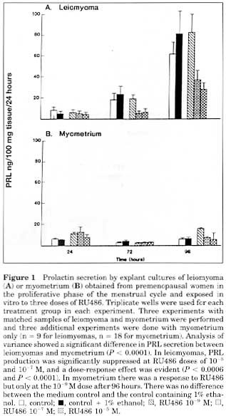
Figure 1.
Figure 2.
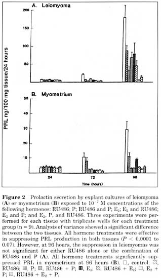
Figure 3.
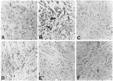
Immunohistochemical staining of leiomyomas (A to C) and normal myometrium (D to F) for PR subtypes (magnification in all cases X178). Sections were incubated with the following primary antibodies: (A and D) 10 mg/mL of nonspecific mouse IgG as a negative control; (B and E) 10 mg/mL of AB-52, which recognizes both A and B isoforms; (C and F) 50 microgram/mL of B-30, which recognizes the B isoform. Staining with B-30 yielded similar results at concentrations of 10 and 20 microgram/mL. Immunostaining of samples from four additional patients were similar. These photographs demonstrate that staining for PR is greater in leiomyomas than in myometrium (B versus E). Both nuclear staining (curved arrow) and cytoplasmic staining (straight arrow) are increased in leiomyomas (B). In addition, for both leiomyomas and myometrium, there is little specific staining for the B isoform (C versus B) and (F versus E). In F, the arrow indicates a cell with no staining except for hematoxylin counterstaining of the nucleus.